颈动脉斑块内出血与临床事件
2018-04-02 16:58:53 by admin ![]() 3739
3739
1. Symptomatic Patients With Mild and Moderate Carotid Stenosis Plaque Features at MRI and Association With Cardiovascular Risk Factors and Statin Use
中等度狭窄患者斑块内出血发生率更高,年龄增长,斑块内出血 几率更高
2. Magnetic Resonance Imaging of Carotid Atherosclerotic Plaque in Clinically Suspected Acute Transient Ischemic Attack and Acute Ischemic Stroke
VI型斑块与同侧急性TIA/IS发作相关,是事件发生的独立标志特征
3. Association Between Carotid Atherosclerosis Plaque With High Signal Intensity on T1-Weighted Imaging and Subsequent Ipsilateral Ischemic Events
患有颈动脉斑块并伴有斑块内出血再发脑梗塞的风险更高
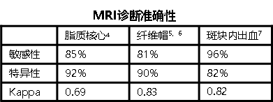
4. In vivo accuracy of multipectral magnetic resonance imaging for identifying lipid-rich necrotic cores and intraplaque hemorrhage in advanced human carotid plaques
5. Visualization of fibrous cap thickness and rupture in human atherosclerotic carotid plaque in vivo with high-resolution magnetic resonance imaging
6. In vivo accuracy of multisequence MR imaging for identifying unstable fibrous caps in advanced human carotid plaques
7. Differentiation of intraplaque versus juxtaluminal hemorrhage/thrombus in advanced human carotid atherosclerotic lesions by in vivo magnetic resonance imaging
结论:
1.斑块内成分(出血)的出现与急性脑梗之间具有显著相关性阴性预测值较高
2.斑块内出血的面积及其与管腔表面的距离潜在的成为判断急性脑梗的影像学指标
3.MRI诊断斑块准确性


 京公网安备 11010502042883号